Protein Crystallization Chip
Dynamic counter-diffusion and dialysis crystallization screening across an integrated clamped semi-permeable membrane.

| Material | 3D-printed polymer, cellulose hydrate membrane, PMMA, anti-evaporation tape, transfer adhesive |
| Method | Mechanically clamped 3D-printed blocks over dialysis membrane, with lower 500 µm channel sealed via laminated optical PMMA base |
| Min feature | 500 µm channel width |
| Idea to tested device | 4 days |
THE WORKFLOW
Crystallization screening is traditionally slow, consumes large volumes of scarce target protein, and relies on static well plates where chemical conditions cannot be adjusted mid-run. Dialysis-based microfluidics decouples the protein sample from a continuously flowing precipitant, allowing dynamic condition gradients without manual pipetting.
HOW IT WAS BUILT
The consumable is built as a hybrid 5-layer assembly. The upper conical microwell block and the microfluidic flow cell were fabricated via high-resolution SLA 3D printing. A cellulose hydrate dialysis membrane is mechanically clamped between the two blocks, creating an immediate, leak-free seal without liquid adhesives that could foul or clog the active pore matrix.
The lower 3D-printed flow cell contains a 500 µm wide fluidic channel with integrated fluidic inlet and outlet. To provide optical clarity for bottom-view inspection, the open lower channel is sealed by laminating an optically clear PMMA base plate using a transfer adhesive. Finally, the upper microwells are sealed with an anti-evaporation tape to minimise vapor loss over hours of continuous screening.
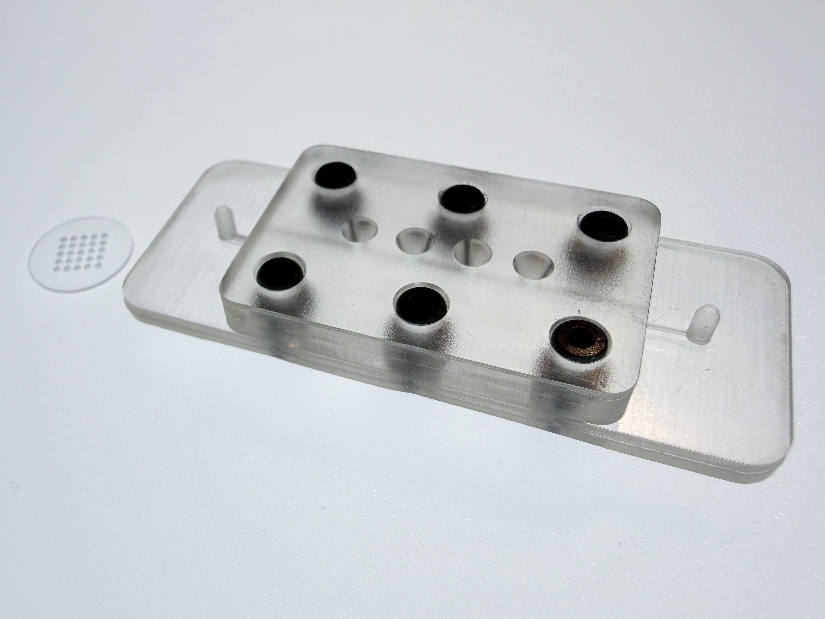 Assembled 3D-printed crystallization cartridge alongside the 10 mm signature glass disc: upper conical microwell block mechanically clamped over the dialysis membrane onto the lower perfusion flow cell with an optically clear PMMA base.
BENCH CHARACTERIZATION
Validated using a lysozyme and NaCl model counter-diffusion system. Dynamic exchange across the cellulose hydrate membrane drives gradual supersaturation inside the microwells. Birefringent protein crystals were successfully grown and optically verified through the bottom viewing window under cross-polarised microscopy within hours.
HOW THIS APPLIES TO YOUR DEVICE
If your workflow requires dynamic dialysis, barrier tissue models, or tangential flow filtration (TFF), integrating delicate semi-permeable membranes into rigid cartridges is notoriously prone to glue fouling and leakage. This architecture proves that membranes can be mechanically clamped and sealed with anti-evaporation tapes in days. The geometry, chamber depths, well counts, and optical inspection windows can be tailored in biocompatible PMMA or 3D resins to match your exact assay protocol and microscope stage.